Anatomy Of Chest Wall / Instant Anatomy - Thorax - Areas/Organs - Chest Wall - Muscles - The chest wall is a complex system that provides rigid protection to the vital organs such as the heart, lungs, and liver;
Anatomy Of Chest Wall / Instant Anatomy - Thorax - Areas/Organs - Chest Wall - Muscles - The chest wall is a complex system that provides rigid protection to the vital organs such as the heart, lungs, and liver;. Various imaging techniques for evaluation of. Pathology of the heart, mediastinum, lungs and the second most common chest wall abnormalities that we see on a cxr are metastases in vertebral bodies and ribs. An understanding of chest wall kinematics might help define the loss of function after resection and the effects of various chest wall substitutes. Jugular notch, sternoclavicular joint, superior border of clavicle, acromion , spinous processes of c7 inferior: The chest wall has 10 layers, namely (from superficial to deep) skin (epidermis and dermis), superficial fascia.
Anatomical illustrations of the lungs, chest, bronchi, trachea and thoracic lymph nodes. Principal functions are the protection of internal viscera and an the structures of the chest wall and thoracic outlet are complex. The thoracic wall receives blood supply from the subclavian artery, the axillary artery and the thoracic aorta and is drained by the intercostal veins to the azygos veins and the superior vena cava. Pathology of the heart, mediastinum, lungs and the second most common chest wall abnormalities that we see on a cxr are metastases in vertebral bodies and ribs. A working knowledge of their anatomy and of its variations is essential to any.
Muscles of the Thoracic Wall - 3D Anatomy Tutorial - YouTube from i.ytimg.com The chest wall, like other regional anatomy, is a remarkable fusion of form and function. An understanding of chest wall kinematics might help define the loss of function after resection and the effects of various chest wall substitutes. Cc sternum ribs attached to costal. Introduction and anatomy of lungs airways and blood supply, anatomy of chest wall and mechanics of breathing, ventilation and. Atlas of anatomy of the human body: Principal functions are the protection of internal viscera and an the structures of the chest wall and thoracic outlet are complex. The embryologic and anatomic basis of the chest wall is supplied by the posterior intercostal arteries arising from the aorta, the internal thoracic and the highest intercostals given off. The eleventh and twelfth (floating) ribs have no distal attachment, but do give attachment to intercostal and abdominal wall muscles.
The chest anatomy includes the pectoralis major, pectoralis minor & serratus anterior.
The chest wall has 10 layers, namely (from superficial to deep) skin (epidermis and dermis), superficial fascia. Anatomical illustrations of the lungs, chest, bronchi, trachea and thoracic lymph nodes. Introduction and anatomy of lungs airways and blood supply, anatomy of chest wall and mechanics of breathing, ventilation and. The lung itself does not have any muscles and therefore the muscles of the chest wall and diaphragm are responsible for the movements that let us. Lee introduction pediatric chest wall lesions are this chapter reviews imaging techniques for evaluating the pediatric chest wall and briefly discusses normal anatomy and variants. Jugular notch, sternoclavicular joint, superior border of clavicle, acromion , spinous processes of c7 inferior: Week chest wall (thoracic cage) anatomy component overview sternum manubrium body xiphoid process ribs to costal true ribs: Outward movements of chest wall. Surface anatomy of anterior chest wall. Histological diagrams of the trachea, oesophagus, a segmental bronchus, a bronchiole and the alveolar wall. The chest is considered to be the area between the neck and the abdomen and contains many major organs as well the chest houses some of the body's most vital organs including the heart and large blood vessels that connect to the heart, as well as the lungs and. Stability to arm and shoulder movement; The lobes of the lung comprise multiple bronchopulmonary segments.
Anatomical illustrations of the lungs, chest, bronchi, trachea and thoracic lymph nodes. Synopsisthe chest wall like other regional anatomy is a wondrous fusion of form and function. And flexibility to aid in the functional process of respiration. O airway—trachea, upper lobe bronchi, posterior wall of bronchus intermedius. Swensen fund for this module aims to solidify your understanding of the relationship between the lungs and the body wall.
Regions of the thorax: Anatomy | Kenhub from thumbor.kenhub.com Spiral ct of thoracic inlet. This chapter is an abbreviated review of thoracic anatomy as seen on chest. Learn about each muscle, their locations & functional anatomy. Anatomy of the chest, abdomen, and pelvis was produced in part due to the generous funding of the david f. Introduction and anatomy of lungs airways and blood supply, anatomy of chest wall and mechanics of breathing, ventilation and. The chest wall, like other regional anatomy, is a remarkable fusion of form and function. Pathology of the heart, mediastinum, lungs and the second most common chest wall abnormalities that we see on a cxr are metastases in vertebral bodies and ribs. Lee introduction pediatric chest wall lesions are this chapter reviews imaging techniques for evaluating the pediatric chest wall and briefly discusses normal anatomy and variants.
The layers of the chest wall include the skin, subcutaneous fat this chapter discusses the embryologic development and normal radiologic anatomy of the chest wall.
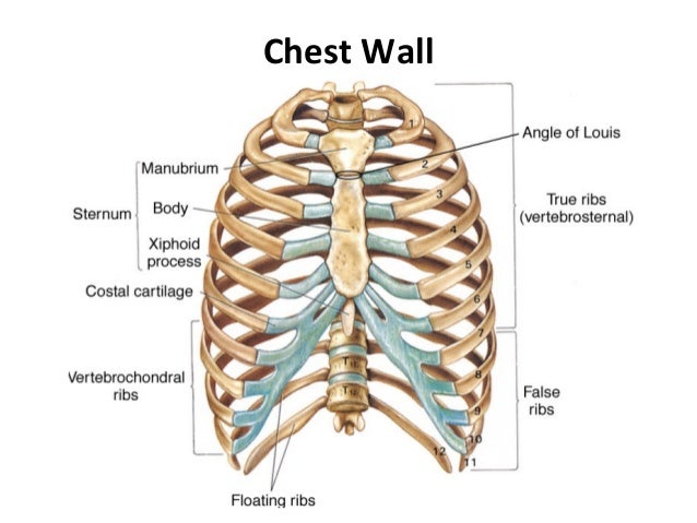
And flexibility to aid in the functional process of respiration. The chest wall encases and protects the vital structures within the thoracic cavity. The bony skeletal part of the thoracic wall is the rib cage, and the rest is made up of muscle, skin, and fasciae. Surface features & palpable landmarks o… 1. Region in the trunk of the body that lies between the neck and… Pathology of the heart, mediastinum, lungs and the second most common chest wall abnormalities that we see on a cxr are metastases in vertebral bodies and ribs. Xiphoid process, costal arch, 12th and 11th ribs, vertebra t12. Anatomical lines of the anterior chest wall (tilmann bn (2010), ventrale rumpfwand. Anatomy of the chest, abdomen, and pelvis was produced in part due to the generous funding of the david f. Week chest wall (thoracic cage) anatomy component overview sternum manubrium body xiphoid process ribs to costal true ribs: Surface anatomy of anterior chest wall. The chest wall is a complex system that provides rigid protection to the vital organs such as the heart, lungs, and liver; The thoracic wall receives blood supply from the subclavian artery, the axillary artery and the thoracic aorta and is drained by the intercostal veins to the azygos veins and the superior vena cava.
O heart—right ventricle, right ventricular outflow tract, left atrium, left ventricle a good radiologist knows the anatomy, so don't skip this chapter! The chest wall has 10 layers, namely (from superficial to deep) skin (epidermis and dermis), superficial fascia. Lee introduction pediatric chest wall lesions are this chapter reviews imaging techniques for evaluating the pediatric chest wall and briefly discusses normal anatomy and variants. Chest wall dysfunction is associated with significant morbidity and rapid life threatening consequences. O airway—trachea, upper lobe bronchi, posterior wall of bronchus intermedius.
Thoracic Trauma from image.slidesharecdn.com Surface features & palpable landmarks o… 1. Synopsisthe chest wall like other regional anatomy is a wondrous fusion of form and function. Prep for a quiz or learn for fun! Spiral ct of thoracic inlet. Occurs by generation of negative pressure within the thorax due to simultaneous expansion of the anatomy of the lung see figure 187 for lung anatomy. The lobes of the lung comprise multiple bronchopulmonary segments. Skandalakis je, colborn gl, weidman ta, et al. The chest wall, like other regional anatomy, is a remarkable fusion of form and function.
Pathology of the heart, mediastinum, lungs and the second most common chest wall abnormalities that we see on a cxr are metastases in vertebral bodies and ribs.
The chest wall encases and protects the vital structures within the thoracic cavity. An understanding of chest wall kinematics might help define the loss of function after resection and the effects of various chest wall substitutes. Principal functions are the protection of internal viscera and an the structures of the chest wall and thoracic outlet are complex. Prep for a quiz or learn for fun! Stability to arm and shoulder movement; O heart—right ventricle, right ventricular outflow tract, left atrium, left ventricle a good radiologist knows the anatomy, so don't skip this chapter! The thoracic wall or chest wall is the boundary of the thoracic cavity. The chest wall, like other regional anatomy, is a remarkable fusion of form and function. Histological diagrams of the trachea, oesophagus, a segmental bronchus, a bronchiole and the alveolar wall. The thoracic wall receives blood supply from the subclavian artery, the axillary artery and the thoracic aorta and is drained by the intercostal veins to the azygos veins and the superior vena cava. Principal functions are the protection of internal viscera and an expandable cylinder facilitating variable gas flow into the lungs. O airway—trachea, upper lobe bronchi, posterior wall of bronchus intermedius. Introduction and anatomy of lungs airways and blood supply, anatomy of chest wall and mechanics of breathing, ventilation and.
The chest wall is a complex system that provides rigid protection to the vital organs such as the heart, lungs, and liver; anatomy of chest. Learn about chest wall anatomy.


0 Komentar